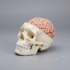
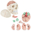
Human Skull & Brain Anatomy Model with Removable Parts (real size)
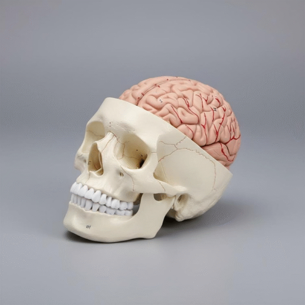
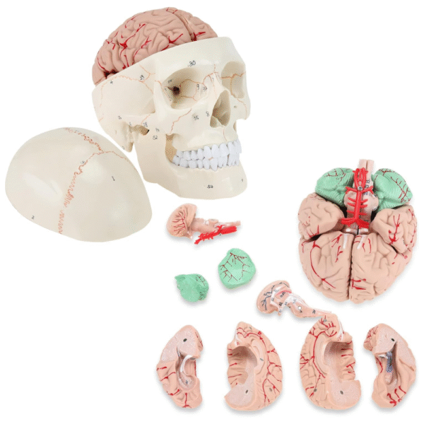
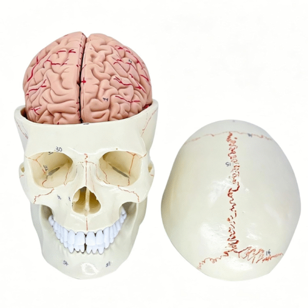
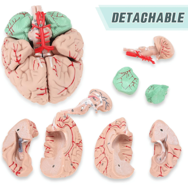
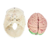
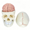
The Human Skull & Brain Anatomy Model is an educational anatomical model designed to demonstrate the structure of the human skull and brain. Its detachable components allow students and healthcare professionals to examine internal brain structures and understand the relationship between different anatomical regions.
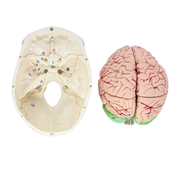
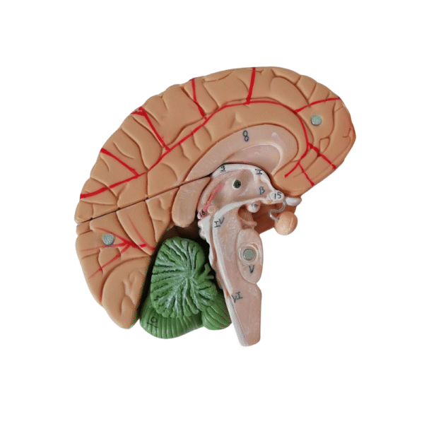
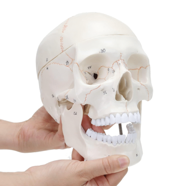
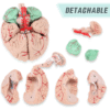
The model features color-coded brain structures, illustrated blood vessels, and numbered anatomical markings for easier identification during teaching and demonstrations.
Anatomical Structures Displayed
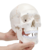
- Human skull: Cranial bones, facial structures, eye sockets, and teeth
- Cerebral hemispheres: Left and right sections of the cerebrum
- Cerebral cortex: Surface folds and grooves of the brain
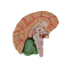
- Cerebellum: Detailed cerebellar structures
- Brainstem: Lower brain structures connecting toward the spinal cord
- Cerebral blood vessels: Illustrated vascular structures across the brain
Key Features
- Detachable skull and brain components
- Detailed representation of brain anatomy
- Includes cerebrum, cerebellum, and brainstem
- Illustrated cerebral blood vessels
- Numbered anatomical markings
- Suitable for medical education and demonstrations
Frequently Asked Questions
Is the skull and brain anatomy model detachable?
Yes. The model includes removable skull and brain sections for studying internal anatomical structures.
What anatomical structures does the model show?
It displays the skull, cerebral hemispheres, cerebellum, brainstem, and illustrated cerebral blood vessels.
Is this model suitable for medical students?
Yes. It is suitable for medical schools, anatomy classrooms, healthcare training, and educational demonstrations.