
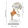
Human Hip Joint Anatomy Model with Ligaments
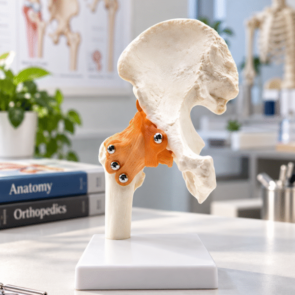
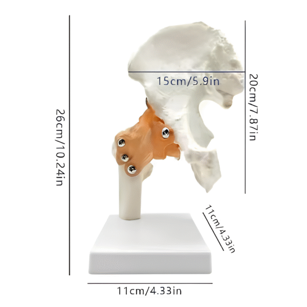
The Human Hip Joint Anatomy Model provides a detailed representation of the hip joint, showing the relationship between the pelvic bone, femoral head, and surrounding ligaments. It helps medical students and healthcare professionals understand hip joint anatomy and the structures responsible for joint stability.
Designed for educational purposes, this anatomical model is suitable for medical schools, physiotherapy clinics, rehabilitation centers, and classrooms.
Key Features
- Anatomical representation of the human hip joint
- Displays the pelvis, femur, and supporting ligaments
- Illustrates hip joint structure and articulation
- Mounted on a stable display base
- Suitable for medical education and demonstrations
Frequently Asked Questions
What does the hip joint anatomy model show?
The model displays the pelvic bone, proximal femur, and supporting ligaments of the human hip joint.
Is this hip anatomy model suitable for medical students?
Yes. It is suitable for anatomy education, medical training, and physiotherapy demonstrations.
Does the hip joint model include a display stand?
Yes. The model comes mounted on a stable base for convenient display.